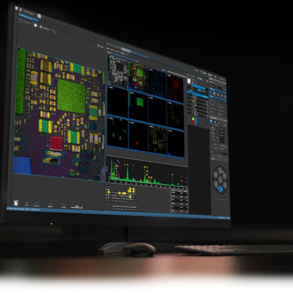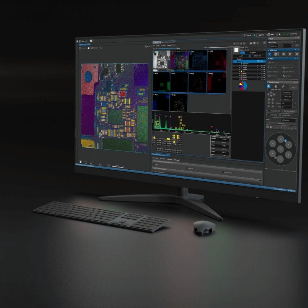

Faster, Ultra-High-Resolution Data
Thanks to Dual Essence™ EDS and Extended Resolution technology, users gain access to fast, high-resolution imaging and compositional data- shortening the time from acquisition to insight.

Efficient
Sample Analysis
With Wide Field Optics™, navigating across large sample surfaces becomes significantly faster—cutting time-to-data by as much as 30% and enabling users to reach critical regions effortlessly.

Seamless Handling of Complex Samples
MultiVac™ mode allows for high-quality imaging of challenging samples—including those that are delicate, outgassing, or non-conductive—without the need for conductive coatings.

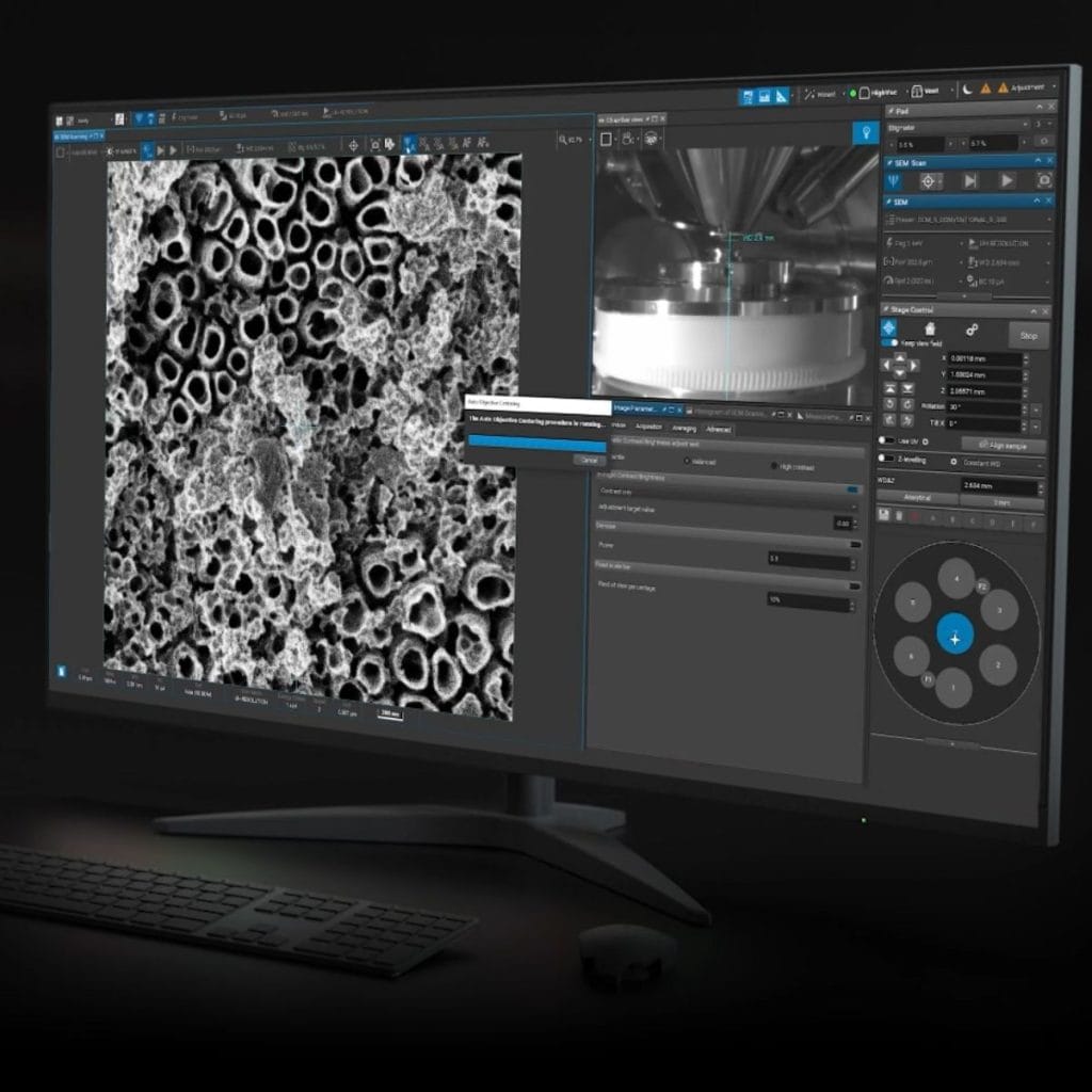
Smart
Automation
In-Flight™ beam optimization and intuitive Essence™ software simplify even the most advanced SEM tasks, allowing users of all experience levels to operate with confidence and consistency.